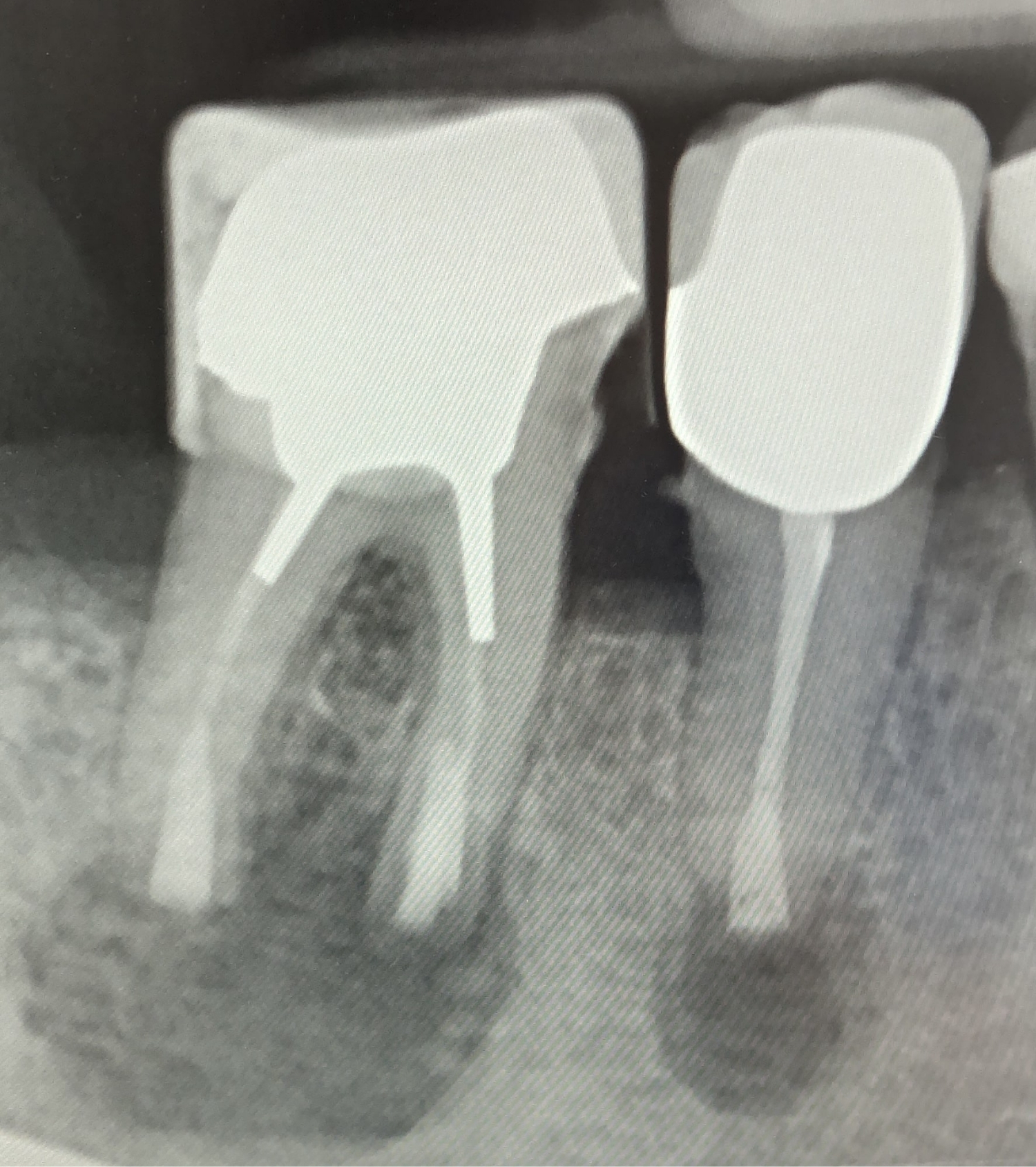
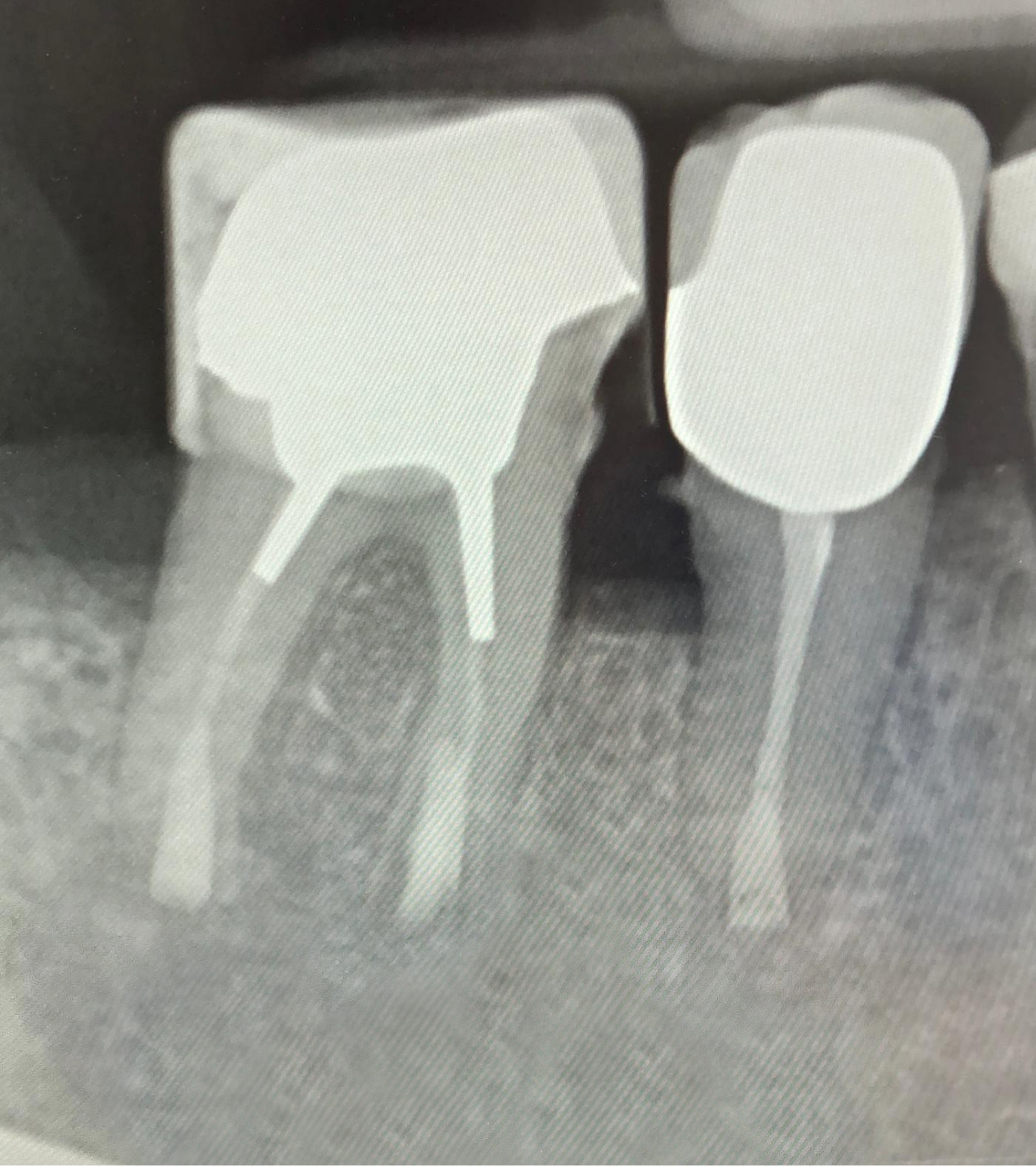
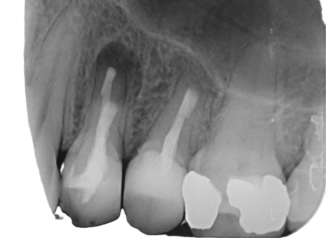
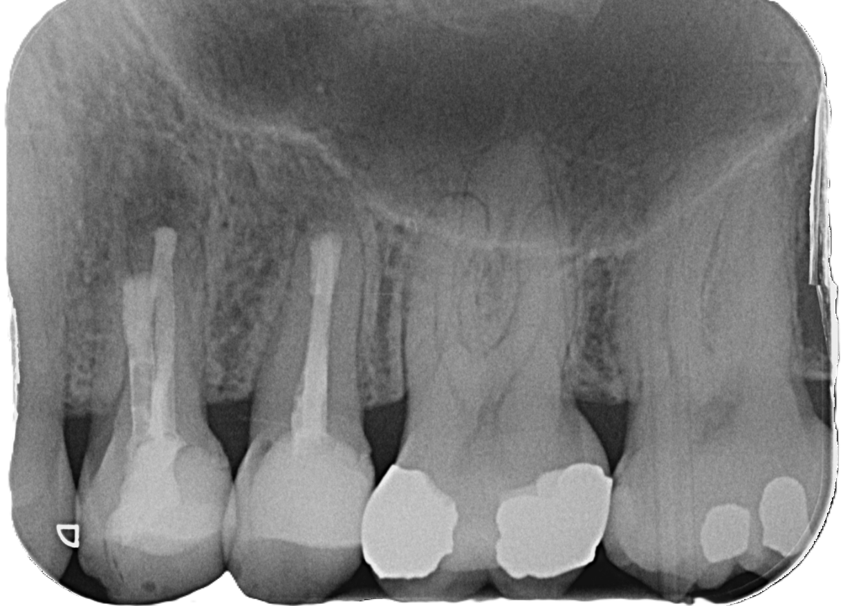
Microsurgery

Microsurgical root tip resection

Microsurgery

Microsurgical root tip resection

Preserving natural teeth – instead of removing them prematurely:

Many patients decide too quickly to have a tooth removed when inflammation occurs in the area of the root tip. However, there are now modern, gentle procedures that can preserve the tooth in the long term.

One of the most successful methods is microsurgical root tip resection (MWR) – a highly precise procedure that saves the natural tooth in most cases.

Zwei digitale Röntgenaufnahmen von Backenzähnen auf einem Monitor. Die Bilder zeigen Zahnwurzeln, Wurzelkanäle und den umgebenden Kieferknochen zur Beurteilung des Zahnzustands.

Before

After

Why not just extract it?

Tooth loss is always a significant disruption to the natural function of the dentition. Replacement with implants or bridges is more costly, more complex, and not always necessary.

After a classic root canal treatment, discomfort may persist. This is due to the complex, individually branched root canal system: not all bacteria can be completely removed.

The result is persistent inflammation in the area of the root tip – associated with pain, swelling, or chronic discomfort. Many patients then see extraction as the only option.

Vorher
Nachher
Zahnärztliches Röntgenbild mit mehreren Backenzähnen – Darstellung von Wurzelkanalbehandlungen und Füllungen zur Diagnostik in der Zahnklinik Nordhorn
Vorher
Zahnärztliches Röntgenbild mit mehreren Backenzähnen – Darstellung von Wurzelkanalbehandlungen und Füllungen zur Diagnostik in der Zahnklinik Nordhorn
Nachher

Microsurgical root tip resection

In MWR, the inflamed tissue at the root tip is microsurgically removed. Using magnifying glasses and an operating microscope, the root canals can then be thoroughly cleaned and sealed with biocompatible materials to prevent bacterial leakage – significantly more effective than with classic methods.

The procedure is based on the microsurgical techniques of Prof. Kim (University of Pennsylvania, USA). Characteristic features include minimally invasive access, the exact removal of only a few millimeters of the affected root tip, high-resolution imaging under up to 40x magnification, and retrograde preparation of the canals. The closure is performed with biocompatible materials that ensure a tight seal and enable complete bone healing.

The result is the permanent elimination of inflammation and the preservation of the natural tooth.

When is MWR useful?

A microsurgical root tip resection may be indicated if:

  • persistent pain occurs when chewing (bite discomfort)
  • the gums in the area of the root tip are swollen or sensitive to pressure
  • a fistula has formed with fluid discharge
  • radiological changes are visible in the area of the root tip
  • a root filling is too short or too long
  • a conventional root tip resection was unsuccessful
  • recurrent infections or an increased susceptibility to infection are present

Success rate in comparison

The classic root tip resection achieves a success rate of only about 40%. With the microsurgical technique according to Prof. Kim, the success rate is over 90%.

The reason for this is the combination of state-of-the-art microscope technology, precise retrograde preparation, and bacteria-tight closure. This allows even the finest lateral canals or micro-cracks to be detected and reliably treated.

Advantages of the microsurgical method

  • Success rate over 90%
  • usually fully capable of working again after 2–3 days
  • complete bone regeneration possible
  • sustainable and more cost-effective alternative to dental prosthetics such as implants or bridges

Success monitoring

To reliably assess the long-term benefits of a therapy, we conduct systematic follow-up examinations. Patients who have been treated with this method usually present again after three months. If necessary, a further check is carried out after six months. In this way, it can be clearly determined whether complete healing has occurred at the root tip.

The result is precisely checked by X-rays: newly formed bone in the area of the root tip indicates that the chewing function has been fully restored. At the same time, patients report on their experiences during the healing process and provide information on the resilience of the affected tooth.

Only when both the radiological findings and the subjective feedback are convincing do we speak of a stable and lasting treatment success. In this case, after our approval, the teeth can be provided by the dentist with dental prosthetics such as crowns, bridges or denture elements.